In 1984, Mehrsheed Sinaki published a study at the Mayo Clinic that should be required reading for every person diagnosed with osteoporosis. It has been cited thousands of times and is still, forty years later, routinely ignored in practice.
She took postmenopausal women with osteoporosis and put them into exercise groups. One group did flexion exercises — sit-ups, toe touches, curling the spine forward. One group did extension exercises — strengthening the muscles that hold the spine upright. One did both. One did neither.
Then she followed them and counted new vertebral compression fractures.
The flexion group fared dramatically worse than the extension group. Loaded, repeated forward bending of an osteoporotic spine is not neutral. It is a fracture mechanism.
What a vertebral compression fracture is
A vertebral compression fracture goes by several names: osteoporotic vertebral fracture, vertebral fragility fracture, crush fracture, wedge fracture, spinal compression fracture, or simply a collapsed vertebra.
The front of the vertebral body — the weight-bearing column — collapses. The back stays intact. The vertebra becomes wedge-shaped. This tips the spine forward, which loads the front of the next vertebra more heavily, which makes the next fracture more likely.
This is why vertebral compression fractures cluster. One fracture substantially raises the risk of the next, and the mechanism is largely mechanical: increasing forward posture increases the compressive load on the anterior column.
Roughly two-thirds are thought to occur without a clear traumatic event. People often describe them as “I just bent over to pick something up.”
The advice that makes it worse
After a diagnosis of osteoporosis or a compression fracture, people are commonly told some version of: be careful, don’t lift, don’t do anything that jars your spine, avoid heavy things.
Follow that advice faithfully and here is what happens over two years:
- Spinal extensor muscles atrophy from disuse
- Bone density declines further from reduced mechanical loading
- Forward posture increases as extensors weaken
- The anterior compressive load rises
- Balance deteriorates, and forward posture shifts the centre of mass
- The next fracture becomes more likely, not less
Protection, applied indefinitely, is the fracture pathway.
What actually reduces risk
Two things: stronger back extensors, and better bone.
Back extensor strength. Sinaki’s later work with spinal extension programs showed reduced kyphosis progression and improved balance. Strong extensors hold the spine upright, which reduces anterior compressive load. Practically: prone extension work, loaded carries, hip hinge patterns taught with a neutral spine, and progressive resistance training.
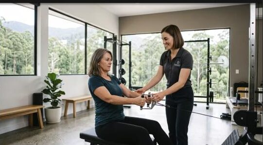
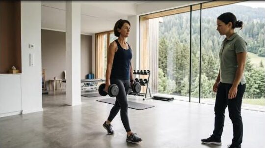
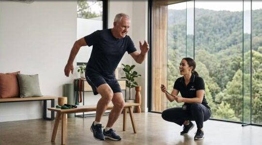
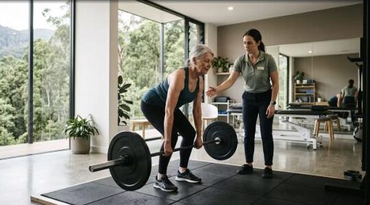
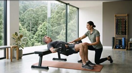
Bone loading. Bone responds to high-magnitude, rapid loading. Walking is good for many things but is a very weak osteogenic stimulus at the spine. Progressive resistance training with meaningful load is a strong one. Australian research — most notably out of Griffith University — has demonstrated that supervised high-intensity resistance and impact training in older adults with low bone mass can improve spine and hip bone density with a strong safety record.
The word doing the work in that sentence is supervised.
What to avoid, honestly
This is where a physio has to be specific rather than vague.
Movements that load the spine into flexion, particularly under load or at speed, carry genuine risk in an osteoporotic spine:
- Sit-ups and crunches
- Loaded toe touches and rounded-back lifting
- Deep forward-bending yoga positions held under load
- Twisting under load, especially combined with flexion
- Certain Pilates spinal articulation drills
This is not “never bend forward.” Life requires bending. It is: don’t repeatedly load a fragile anterior vertebral column into flexion, and learn to hinge from the hip so that heavy tasks travel through a neutral spine.
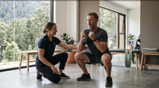
Teaching a proper hip hinge is one of the highest-value things we do at Longevity Physio Studio in Eagle Heights, and it takes most people about three sessions to own it.
The longevity picture
Vertebral compression fractures are associated with increased mortality, and — as with hip fractures — the mechanism is largely downstream: pain, reduced activity, reduced lung capacity from progressive kyphosis, reduced appetite, deconditioning, falls.
Height loss is a signal worth taking seriously. If you have lost more than 3 to 4 cm from your peak adult height, that warrants investigation.
What an assessment involves
At our Eagle Heights clinic on Tamborine Mountain, an osteoporosis and spine assessment includes height measurement against known peak height, thoracic kyphosis assessment, back extensor endurance testing, hip hinge competence, balance testing, grip strength, and a review of your DEXA results if you have them.
Then we build a program that loads the spine safely rather than avoiding it indefinitely.
Book an assessment
If you have osteoporosis, osteopenia, a past compression fracture, or you have lost height, the question is not whether to load your spine. It is how.
Longevity Physio Studio, Eagle Heights, Mt Tamborine — serving Tamborine Mountain, North Tamborine, Canungra and the Scenic Rim. Book an assessment.